
A radiographer produces images of specific parts of the body through the use of radiology equipment. There are different pieces of equipment for producing different, specialized images. Some areas of specialty include mammography, computed tomography (CT) scanning, and X-ray. While some radiographers choose an area of specialty, many work in settings that allow the individual to produce images for a variety of purposes. All of these machines use radiation, thus safety precautions must always be taken to protect both the patient and the radiographer. Whether you choose to specialize or not, your school's program will introduce you to most of the equipment that you may encounter during your career.
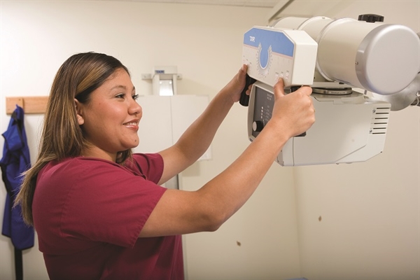
The first imaging device that comes to mind for many is the simple X-ray machine. Invented during the late 1800s, this machine uses a single beam of radiation to create an image on film, similar to what a camera does. These machines may be large and contained within a designated room, or they may be in portable format and used when a patient cannot be transported to a different area for imaging studies. The X-ray images are produced on large pieces of film that can be viewed almost immediately.
Perhaps one of the most interesting pieces of radiology equipment is the fluoroscope. The fluoroscope works in much the same way that an X-ray machine does, but it includes a screen that allows the image to be seen in real time. For instance, in a clinical setting, an orthopedist will use this instrument when "re-setting" a broken bone in order to visualize whether the bone is in the correct position before the patient leaves the office.
A CT, or computed tomography scanner, is a very large piece of equipment. It is generally a cylinder, and the patient lies on a table that slides into the machine. It uses multiple beams of radiation to create a two-dimensional picture of structures inside the body, transferring these images onto a computer screen. Often, contrast media such as barium is used in CT studies.
Another very common piece of radiology equipment with which most women will come into contact during their lives is the mammography machine. This device is designed to be used with the patient standing, and as with most other X-rays, the radiographer manipulates the body part — in this case, the breast — in order to image it from different angles. Although this machine uses radiation to create its images, the newer devices capture digital images rather than using film.
It is always the radiographer's goal to use the least amount of radiation necessary to produce a quality image that will serve its purpose in the patient's diagnosis and care. The radiographer is responsible for not only the patient's safety and documenting the amount of radiation used, but they are also responsible for his or her own safety and protection. In this career, you may be in charge of maintaining and troubleshooting these machines, but your coursework will prepare you for these tasks.
With the U.S. Bureau of Labor Statistics predicting 28 percent job growth in the career field of radiologic technology, now is a good time to enter this profession if you think that it may be right for you.