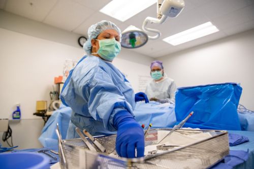
Just about everyone has some type of imaging done on their body, with X-rays being the most simple and common procedure available. Radiologic technology is being used by doctors now more than ever because of the dynamic changes in technology, machinery and quick results. These continually improving trends make our list of the top three advancements in radiology that you may not tackle in the classroom.
Instant Gratification
Most often, patients ask when their doctor will receive results of tests that were done on a visit. In our fast-paced world in which the words "digital" and "download" are common vocabulary, the trend is moving toward sharing downloadable images between doctor and clinic to make diagnosing faster, more efficient and less costly. Radiologists now analyze images on computer screens and dictate reports on sophisticated voice recognition software rather than hanging bulky films on light boxes and waiting for transcriptionists to do the report typing. Results typically reach the ordering physician within 24 hours, perhaps even on the same day, which is a great improvement in turnaround time from just a few years ago. Sharing images digitally is as fast as a few clicks of a mouse. For the sake of portability, a CD of the images can easily be provided to the patient with the software program already in place and set to run on most any computer.
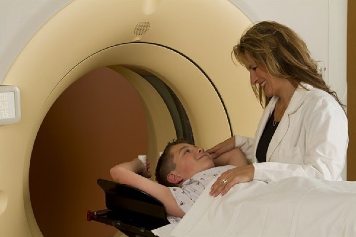
Less Radiation Exposure in CT
Any ability to minimize a patient's exposure to radiation is a benefit. In recent years, CT machinery has improved to create more slices while delivering lower doses of radiation. In the past, 16 slice scanners were the industry standard as compared to present day, a time in which 128 slice, high-definition CT scanners are, slowly but surely, making their way into hospitals and clinics across the country. Thanks to highly improved technology, the 128 slice CT is able to refine and increase image detail, creating sharp 3D images in seconds, while delivering much lower doses of radiation than the older machinery.
Digital Mammograms
Breast cancer is the second most common cancer plaguing women, so the gradual move from film to digital mammography has allowed for a crisper, more accurate picture of the breast tissue, which may better detect the presence of breast cancer due to analysis by a radiologist and by computer-aided detection (CAD). Where films are a permanent picture that can't be changed, a digital picture can easily be manipulated by rotating the image, zooming in on a specific area or changing the contrast of the picture for better viewing. Although film mammograms are still highly effective, advanced digital technology is better able to detect abnormalities in individuals with denser breasts, as both normal tissue and tumors appear white, which can make it difficult to distinguish between typical and atypical breast tissue.
Although these advancements may still be too new to be introduced in a classroom setting, they are just a few of the progressive improvements being made in the field of radiologic technology. This will ultimately be a more effective link and better form of communication between the technologist, radiologist, ordering physician, and most importantly, the patient.