
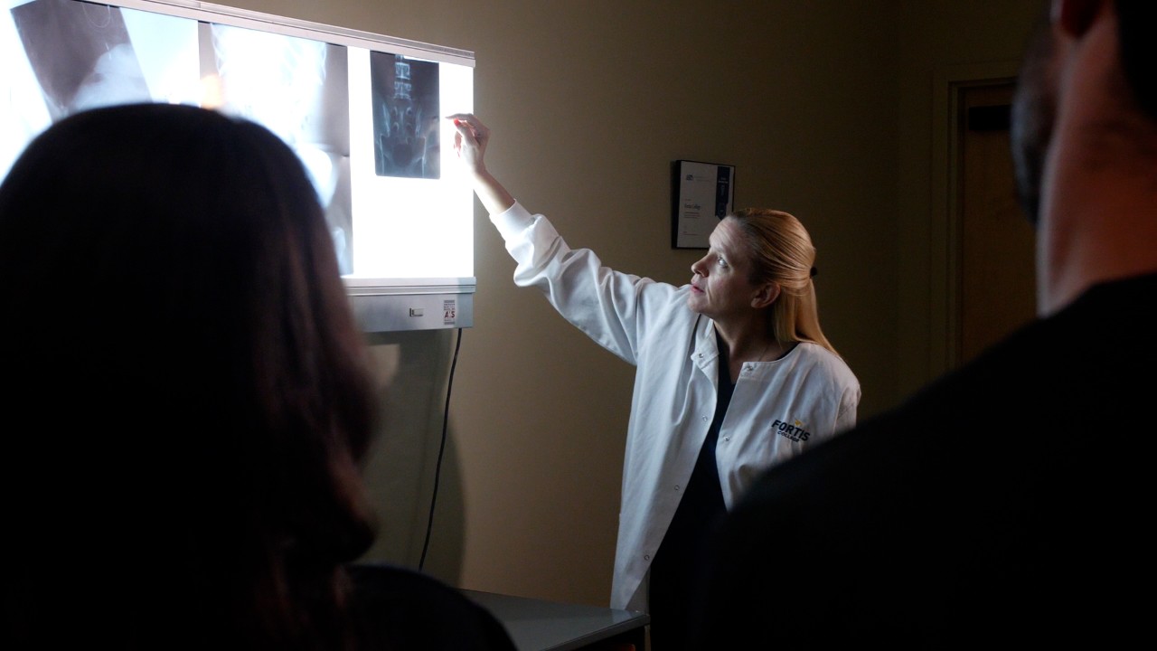
Radiology technologists play a central role on healthcare teams. They are the experts who take images of bones, internal organs, and soft tissue to help physicians diagnose conditions or illnesses. Without imaging to see what’s going on inside the body, doctors might just be guessing about the reason for a patient’s pain. Although technological advances have resulted in a transition from the use of film for image capture to today’s digital imaging, the need for X-rays and their usefulness remains unchanged.
Radiology technologists, aka rad techs, are trained to take different kinds of X-ray exams. While some of these specialties require extra training, here are three types of imaging a rad tech could be in charge of as a member of a healthcare team.
X-ray. Plain X-rays are the mainstay of imaging. The X-ray machine uses ionizing radiation to diagnose things like bone fractures, a swallowed object lodged internally, and arthritis and osteoporosis. X-rays can also identify digestive tract issues and infections.
Mammography is a specialized subset of X-ray technology that uses a special X-ray machine to image breast tissue and screen for cancer. The imaging can find abnormalities that are too small to be detected by physical exam.
To perform an X-ray, the rad tech positions the patient, either lying, sitting, or standing, in front of the X-ray machine to take images. They make take several pictures from different angles. These types of X-rays are short in duration, about 10-15 minutes.
CT Scan. A CT scan, short for “computed tomography,” also uses ionizing radiation to diagnose some of the same issues a plain X-ray captures, as well as soft tissue injuries or conditions. The patient lies on a table that slides into a cylindrical scanner, which rotates around the patient to take a series of X-rays capturing a cross section of the patient’s body. These image slices are stacked to give a complete picture, and allow physicians to see the inside of organs, which can’t be seen by a regular X-ray.
Some of the health issues a CT is used for include bone fractures, injuries from trauma such as a car accident or a fall, infection, cancer, and heart or vascular disease. These X-rays also only take about 10-15 minutes.
MRI scan (magnetic resonance scan). As with a CT scan, a patient slides into a cylindrical scanner for an MRI, but the scanner is thinner and deeper, and it includes loud popping sounds as the scanner captures images. Using magnetic fields and radio waves, MRIs are used to image the condition of soft tissue, including tendon and ligament injuries, tumors, spinal injuries or disease, and organs (brain, heart, abdominal organs, etc.). MRIs can’t X-ray bones because bones don’t contain water the way soft tissue does. These scans can take significantly longer than a CT scan, sometimes up to an hour.
If X-ray technology sounds interesting to you, Fortis offers an associate degree program that trains students for entry-level positions in radiological technology. Click here to learn more about the program, or call 1-855-436-7847 for more information.